PHYSIOLOGY
?? Complete Textbook · 2025 Edition

Human
Physiology

A comprehensive, evidence-based exploration of how the body functions — from membrane potentials to whole-body exercise responses. With annotated diagrams, educational videos, and multilingual audio.

12Chapters
59Topics
12Videos
??Audio · 8 Langs
???Diagrams
Introduction ??

Introduction to Physiology

The Science of How the Body Works

?? 5 Sections ?? Video ?? Audio ??? Diagram
??? Diagram
Human body systems — the integrated machine of life
```
?? Video
?
Introduction to Physiology
Introduction to Physiology · Educational · YouTube
```
?? Audio: Introduction to Physiology
Click Listen to start audio explanation
```

What Is Physiology?

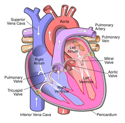
Physiology is the branch of biology and medicine that studies the normal functions of living organisms and their parts. Derived from the Greek physis (nature) and logos (study), physiology asks: How does the heart pump? How do neurons fire? How does the kidney filter blood? It is the foundational science bridging anatomy (structure) with clinical medicine (dysfunction and disease). Physiology is inherently dynamic — it studies processes, not just parts.

Homeostasis: The Central Principle

The most fundamental concept in physiology is homeostasis — the ability of the body to maintain a stable internal environment despite external changes. Claude Bernard (1813–1878) coined the idea of the milieu intérieur (internal environment). Walter Cannon later named it homeostasis. Key variables maintained include body temperature (37°C ± 0.5°C), blood pH (7.35–7.45), blood glucose (70–110 mg/dL), plasma osmolarity (280–295 mOsm/L), and blood pressure (~120/80 mmHg). Disruption of homeostasis is the definition of disease.

Feedback Control Systems

Homeostasis is maintained through feedback loops. Negative feedback loops oppose a change and restore the setpoint — for example, when body temperature rises, sweating and vasodilation cool the body. This is the most common type. Positive feedback amplifies a change — for example, during childbirth, oxytocin causes uterine contractions that press the baby against the cervix, which triggers more oxytocin — a self-amplifying cycle that ends only with birth. Blood clotting and the action potential in neurons are also positive feedback events.

Cell as the Unit of Physiology

All physiological functions ultimately reduce to cellular processes. The cell membrane (phospholipid bilayer with embedded proteins) controls what enters and leaves. Ion channels, pumps (Na?/K?-ATPase), and carriers maintain electrochemical gradients essential for nerve impulses, muscle contraction, and nutrient absorption. Mitochondria produce ATP via oxidative phosphorylation — the energy currency for every active physiological process. Understanding cell physiology is the key to understanding organ physiology.

Levels of Physiological Study

Physiology operates across multiple scales: molecular physiology (protein conformational changes, enzyme kinetics, gene expression), cellular physiology (action potentials, receptor signaling, secretion), organ physiology (cardiac output, lung mechanics, glomerular filtration), systems physiology (cardiovascular regulation, hormonal axes), and whole-organism physiology (exercise response, stress adaptation, aging). Modern physiology integrates all these levels through computational modeling and systems biology approaches.

```
?
Chapter 1 ??

Cell Physiology

Membrane Transport, Potentials & Signaling

?? 5 Sections ?? Video ?? Audio ??? Diagram
??? Diagram
Animal cell ultrastructure showing organelles and membrane systems
```
?? Video
?
Cell Physiology & Membrane Transport
Cell Physiology & Membrane Transport · Educational · YouTube
```
?? Audio: Cell Physiology
Click Listen to start audio explanation
```

The Cell Membrane & Transport

The plasma membrane is a fluid mosaic of phospholipids and proteins. Transport across it occurs via: simple diffusion (O2, CO2, lipid-soluble drugs — down concentration gradient, no energy); facilitated diffusion (glucose via GLUT transporters — needs carrier protein, no energy); osmosis (water movement through aquaporins toward higher solute concentration); active transport (Na?/K?-ATPase pumps 3 Na? out and 2 K? in per ATP — creating the electrochemical gradient that powers most other transport). Secondary active transport uses this gradient to co-transport other solutes.

Membrane Potential & The Resting Potential

All cells maintain a resting membrane potential — a voltage across the plasma membrane. In most neurons and muscle cells, this is approximately -70 mV (inside negative). This potential arises because K? ions diffuse out through leak channels (the membrane is more permeable to K? at rest), and the Na?/K?-ATPase actively maintains the ionic asymmetry. The Nernst equation calculates the equilibrium potential for each ion; the Goldman equation integrates multiple ions to give the actual resting potential.

The Action Potential

When a neuron or muscle cell is depolarized to threshold (~-55 mV), voltage-gated Na? channels open explosively — Na? rushes in, causing rapid depolarization to +30 mV (the peak). This is the upstroke. Then Na? channels inactivate while voltage-gated K? channels open — K? rushes out, repolarizing the membrane. Brief hyperpolarization (afterhyperpolarization) follows before resting potential is restored. The refractory period (absolute: no new AP possible; relative: requires stronger stimulus) ensures unidirectional propagation along the axon.

Cell Signaling & Receptors

Cells communicate via ligands binding to receptors. Ion channel-linked receptors (ionotropic) directly open ion channels — fast, milliseconds (nicotinic acetylcholine receptor). G-protein coupled receptors (GPCRs) activate second messengers: Gs stimulates adenylyl cyclase ? cAMP ? PKA (epinephrine on heart); Gi inhibits adenylyl cyclase; Gq activates phospholipase C ? IP3 + DAG ? Ca²? release + PKC. Enzyme-linked receptors (tyrosine kinases) mediate growth factor responses. Nuclear receptors bind steroid hormones and regulate gene transcription.

Endocytosis, Exocytosis & Vesicular Transport

Cells import large molecules via endocytosis: phagocytosis (engulfing solid particles — macrophages ingest bacteria), pinocytosis (engulfing fluid), and receptor-mediated endocytosis (LDL uptake via clathrin-coated pits). Exocytosis releases vesicle contents — constitutive secretion (continuous, e.g., mucus) or regulated secretion (triggered by Ca²? influx, e.g., neurotransmitter release at synapses, insulin from beta cells). Synaptic vesicle fusion with the presynaptic membrane exemplifies calcium-triggered, SNARE protein-mediated exocytosis.

```
?
Chapter 2 ?

Neurophysiology

Nerve Impulses, Synapses & Neural Circuits

?? 5 Sections ?? Video ?? Audio ??? Diagram
??? Diagram
Structure of a myelinated neuron with axon terminals
```
?? Video
?
Neurophysiology & Action Potentials
Neurophysiology & Action Potentials · Educational · YouTube
```
?? Audio: Neurophysiology
Click Listen to start audio explanation
```

Saltatory Conduction & Myelination

Unmyelinated axons conduct impulses by continuous depolarization along the membrane — slow (0.5–2 m/s). Myelinated axons (Schwann cells in PNS, oligodendrocytes in CNS) have insulated internodes with exposed nodes of Ranvier. The action potential 'jumps' between nodes — saltatory conduction — reaching speeds of 70–120 m/s. Axon diameter also matters: larger diameter = lower resistance = faster conduction. Multiple sclerosis is an autoimmune demyelinating disease, causing slowed or blocked conduction.

Synaptic Transmission

At a chemical synapse, an action potential arrives at the presynaptic terminal ? voltage-gated Ca²? channels open ? Ca²? influx triggers vesicle fusion (SNARE proteins: synaptobrevin, SNAP-25, syntaxin) ? neurotransmitter released into synaptic cleft ? binds postsynaptic receptors. The signal is terminated by reuptake (serotonin transporter — target of SSRIs), enzymatic degradation (acetylcholinesterase breaks down ACh), or diffusion. Electrical synapses (gap junctions) allow direct ion flow — faster but less modifiable than chemical synapses.

Neurotransmitters & Their Functions

Acetylcholine (ACh): nicotinic receptors (skeletal muscle NMJ, sympathetic ganglia); muscarinic receptors (heart slowing, gland secretion, smooth muscle). Glutamate: main excitatory CNS neurotransmitter — AMPA and NMDA receptors. GABA: main inhibitory CNS neurotransmitter — opens Cl? channels ? hyperpolarization. Dopamine: reward, motor control (substantia nigra?striatum); depletion causes Parkinson's. Serotonin: mood, sleep, appetite. Norepinephrine: arousal, fight-or-flight. Substance P, neuropeptides: modulate pain transmission.

Sensory Physiology & Receptor Potentials

Sensory transduction converts stimuli into receptor potentials. Mechanoreceptors (Meissner, Pacinian, Merkel, Ruffini) respond to touch/pressure; nociceptors to tissue damage (pain); photoreceptors (rods and cones) to light via phototransduction (rhodopsin ? G-protein ? cGMP breakdown ? channel closure ? hyperpolarization — uniquely, light causes hyperpolarization, not depolarization). Proprioceptors (muscle spindles, Golgi tendon organs) sense body position. Stimulus intensity is coded by action potential frequency (rate coding) and number of recruited fibers.

Spinal Cord Physiology & Reflex Arcs

The simplest reflex arc — the monosynaptic stretch reflex — involves only two neurons: the Ia afferent from the muscle spindle synapses directly on the alpha motor neuron. Tapping the patellar tendon stretches the quadriceps ? spindle fires ? monosynaptic excitation of quadriceps motor neuron ? leg kicks. Simultaneously, reciprocal inhibition via interneurons relaxes the hamstrings. Polysynaptic reflexes (withdrawal/flexor reflex) involve interneurons. Spinal reflexes operate independently of the brain but are normally modulated by descending cortical and brainstem pathways.

```
?
Chapter 3 ??

Muscle Physiology

Contraction Mechanisms & Motor Control

?? 5 Sections ?? Video ?? Audio ??? Diagram
??? Diagram
Sarcomere structure showing actin, myosin, and Z-lines
```
?? Video
?
Muscle Physiology & Contraction
Muscle Physiology & Contraction · Educational · YouTube
```
?? Audio: Muscle Physiology
Click Listen to start audio explanation
```

Excitation-Contraction Coupling

The neuromuscular junction (NMJ): ACh released from motor nerve terminal binds nicotinic receptors on the motor end plate ? end plate potential ? action potential propagates along muscle fiber ? enters T-tubules (deep invaginations) ? activates dihydropyridine receptors (DHPR) on T-tubule membrane ? conformational change triggers ryanodine receptors (RyR1) on sarcoplasmic reticulum ? massive Ca²? release into cytoplasm (from ~100 nM to ~10 µM). Ca²? binds troponin C on thin filaments ? conformational change exposes myosin-binding sites on actin ? contraction begins.

The Cross-Bridge Cycle

The cross-bridge cycle drives sarcomere shortening: (1) Myosin head (with ADP+Pi bound) binds actin ? cross-bridge forms. (2) Pi release ? power stroke — myosin head pivots 45°?60°, pulling actin filament ~10 nm toward M-line. ADP released. (3) New ATP binds myosin ? cross-bridge detaches. (4) ATP hydrolysis (ATPase) ? myosin re-cocks to high-energy position. Cycle repeats while Ca²? is elevated. Rigor mortis occurs when ATP is depleted postmortem — myosin heads cannot detach, causing rigid muscles (peaks 12 hours, resolves 72 hours).

Twitch, Summation & Tetanus

A single action potential produces one twitch (brief contraction-relaxation cycle). If a second stimulus arrives before full relaxation, twitches sum (wave summation or temporal summation), producing greater force. With rapid stimulation, Ca²? never fully returns to SR — sustained maximal contraction called tetanus (not to be confused with the disease). Tetanic force is 3–4× single twitch force. In vivo, smooth sustained contractions result from asynchronous recruitment of motor units (spatial summation), so different units take turns — preventing fatigue.

Cardiac Muscle Physiology

Cardiac muscle is striated and involuntary, with unique features. Intercalated discs contain gap junctions (electrical coupling for coordinated contraction) and desmosomes (mechanical coupling). The cardiac action potential has a prolonged plateau (phase 2) due to L-type Ca²? channel opening — Ca²? influx triggers Ca²?-induced Ca²? release (CICR) from SR via RyR2. The long action potential duration (~250 ms) creates a long refractory period preventing tetanus — essential for the heart to refill between beats. Contractility is modulated by sympathetic/parasympathetic tone and Frank-Starling mechanism.

Smooth Muscle Physiology

Smooth muscle lacks troponin and sarcomeres. Contraction is triggered by Ca²? (from SR or extracellular) binding calmodulin ? Ca²?-calmodulin complex activates myosin light chain kinase (MLCK) ? phosphorylates myosin light chains ? ATPase activity and cross-bridge cycling. Relaxation: myosin light chain phosphatase (MLCP) dephosphorylates myosin. Tone is regulated by autonomic nerves, hormones, local metabolites, and stretch (myogenic response). Vascular smooth muscle myogenic response: stretch ? contraction (autoregulation of blood flow).

```
?
Chapter 4 ??

Cardiovascular Physiology

Cardiac Function, Blood Pressure & Regulation

?? 5 Sections ?? Video ?? Audio ??? Diagram
??? Diagram
Heart chambers, valves, and direction of blood flow
```
?? Video
?
Cardiovascular Physiology
Cardiovascular Physiology · Educational · YouTube
```
?? Audio: Cardiovascular Physiology
Click Listen to start audio explanation
```

The Cardiac Cycle & Pressure-Volume Loop

The cardiac cycle has two phases: systole (contraction, ~300 ms) and diastole (relaxation, ~500 ms at 75 bpm). During systole: isovolumetric contraction (both valves closed, pressure rises) ? ejection (aortic valve opens, blood ejected). During diastole: isovolumetric relaxation (all valves closed, pressure falls) ? ventricular filling (AV valves open, passive then active filling). The pressure-volume loop graphically represents this cycle — its area = stroke work. Ejection fraction (EF = SV/EDV × 100) is normally >55%; reduced EF indicates systolic heart failure.

Cardiac Output & Its Determinants

Cardiac output (CO) = Heart Rate (HR) × Stroke Volume (SV). Normal CO at rest: ~5 L/min; during exercise: up to 25 L/min in untrained, 40 L/min in elite athletes. Stroke volume is determined by: preload (EDV — Frank-Starling law: greater stretch ? greater force; regulated by venous return), afterload (aortic pressure — higher afterload reduces SV), and contractility (inotropy — sympathetic stimulation via ß1 receptors increases Ca²? and force). HR is regulated by the SA node, modulated by sympathetic (increases HR) and parasympathetic/vagal (decreases HR) tone.

Blood Pressure Regulation

Mean arterial pressure (MAP) = CO × Total Peripheral Resistance (TPR). Short-term regulation (seconds-minutes): arterial baroreceptors (carotid sinus, aortic arch) detect pressure changes ? reflex adjustment of HR, contractility, and vascular tone via autonomic nervous system. Medium-term: capillary fluid shift, stress relaxation of vessels. Long-term (hours-days): kidney controls blood volume via renin-angiotensin-aldosterone system (RAAS): renin ? angiotensin I ? ACE ? angiotensin II (vasoconstriction + aldosterone release ? Na?/water retention ? increased blood volume ? increased MAP).

Microcirculation & Capillary Exchange

Starling's forces govern fluid movement across capillary walls. Hydrostatic pressure (Pc) pushes fluid out; oncotic pressure (pc, from plasma proteins, mainly albumin) pulls fluid in. Net filtration = Kf [(Pc - Pi) - s(pc - pi)]. At the arterial end, hydrostatic pressure dominates ? fluid filters out. At the venous end, oncotic pressure dominates ? fluid reabsorbed. Approximately 3 L/day net filters out and is returned by lymphatics. Disruption causes edema: heart failure (increased Pc), hypoalbuminemia (decreased pc), lymphatic obstruction.

Special Circulations

Coronary circulation: blood flows to myocardium mainly during diastole (systolic compression compresses coronary vessels). Left coronary flow is nearly absent in systole. Metabolic autoregulation: adenosine (from ATP breakdown) is the primary vasodilator — cardiac ischemia releases adenosine, causing coronary vasodilation. Cerebral circulation: autoregulates blood flow over MAP 60–150 mmHg; CO2 is the primary regulator — hypercapnia ? cerebral vasodilation. Pulmonary circulation: hypoxic vasoconstriction (unique — opposite of systemic) diverts blood from poorly ventilated alveoli to well-ventilated ones.

```
?
Chapter 5 ??

Respiratory Physiology

Ventilation, Gas Exchange & Control of Breathing

?? 5 Sections ?? Video ?? Audio ??? Diagram
??? Diagram
The respiratory system: upper airways to alveoli
```
?? Video
?
Respiratory Physiology
Respiratory Physiology · Educational · YouTube
```
?? Audio: Respiratory Physiology
Click Listen to start audio explanation
```

Lung Mechanics & Compliance

Breathing requires generating pressure differences. Compliance (C = ?V/?P) measures lung distensibility — normal lung compliance is ~200 mL/cmH2O. Reduced compliance (stiff lungs) occurs in pulmonary fibrosis, infant RDS (surfactant deficiency). Increased compliance (floppy lungs) occurs in emphysema. Surface tension at the alveolar air-liquid interface tends to collapse alveoli — pulmonary surfactant (dipalmitoylphosphatidylcholine, produced by type II pneumocytes) reduces surface tension by 10-fold, preventing alveolar collapse and reducing work of breathing. LaPlace's law: P = 2T/r — smaller alveoli would collapse into larger ones without surfactant equalizing surface tension.

Spirometry & Lung Volumes

Key lung volumes: Tidal Volume (TV) = 500 mL (normal breath); Inspiratory Reserve Volume (IRV) = ~3000 mL; Expiratory Reserve Volume (ERV) = ~1200 mL; Residual Volume (RV) = ~1200 mL (cannot be expired). Capacities: Total Lung Capacity (TLC) = ~6000 mL; Functional Residual Capacity (FRC) = ERV + RV = ~2400 mL; Vital Capacity (VC) = IRV + TV + ERV = ~4700 mL. Spirometry distinguishes obstructive (FEV1/FVC <0.7 — asthma, COPD — increased RV, air trapping) from restrictive (reduced TLC, FVC — fibrosis, obesity, neuromuscular disease — normal FEV1/FVC ratio).

Gas Exchange & Oxygen Transport

Alveolar gas exchange follows Fick's law: flux = (surface area × diffusion coefficient × ?P) / membrane thickness. Normal alveolar PO2 = 100 mmHg; arterial PO2 = 95 mmHg. Oxygen is transported: 98.5% bound to hemoglobin (each Hb carries 4 O2; cooperative binding gives S-shaped oxyhemoglobin dissociation curve), 1.5% dissolved. The Bohr effect: increased PCO2, decreased pH, increased temperature, and increased 2,3-BPG right-shift the curve (reduced O2 affinity ? more O2 released to tissues). Fetal Hb (HbF) has left-shifted curve — higher affinity, extracting O2 from maternal blood.

CO2 Transport & Acid-Base

CO2 transport: 7% dissolved, 23% bound to hemoglobin as carbaminohemoglobin (Haldane effect: deoxygenated Hb carries more CO2), 70% as bicarbonate. In red blood cells: CO2 + H2O ? H2CO3 ? H? + HCO3? (catalyzed by carbonic anhydrase). HCO3? exits via Cl?/HCO3? exchanger (chloride shift). Acid-base physiology: pH = 6.1 + log([HCO3?] / 0.03 × PCO2) (Henderson-Hasselbalch). Respiratory acidosis (?PCO2), respiratory alkalosis (?PCO2), metabolic acidosis (?HCO3?), metabolic alkalosis (?HCO3?).

Control of Breathing

The respiratory rhythm generator (pre-Bötzinger complex in medulla) generates the basic breathing rhythm. The dorsal respiratory group (DRG) drives inspiration; ventral respiratory group (VRG) drives forced expiration. Central chemoreceptors on the ventral medullary surface respond to PCO2/H? changes in CSF — the most sensitive (70% of hypercapnic drive). Peripheral chemoreceptors (carotid and aortic bodies) respond primarily to PO2 (<60 mmHg triggers significant response), and also to PCO2 and pH — the hypoxic drive. COPD patients may rely on hypoxic drive — supplemental O2 can dangerously blunt their respiratory drive.

```
?
Chapter 6 ??

Renal Physiology

Filtration, Reabsorption, Secretion & Homeostasis

?? 5 Sections ?? Video ?? Audio ??? Diagram
??? Diagram
Kidney cross-section showing cortex, medulla, and collecting system
```
?? Video
?
Renal Physiology & Kidney Function
Renal Physiology & Kidney Function · Educational · YouTube
```
?? Audio: Renal Physiology
Click Listen to start audio explanation
```

Glomerular Filtration

Glomerular filtration rate (GFR) = 125 mL/min = 180 L/day. The glomerular filtration barrier has three layers: fenestrated capillary endothelium (size barrier), glomerular basement membrane (charge + size barrier — negatively charged, repels albumin), and podocyte slit diaphragms (filtration slits between foot processes). GFR is determined by Starling forces: Net filtration pressure = (Pgc - Pbs) - (pgc - pbs) ˜ +10 mmHg. Filtration fraction = GFR/RPF = 20%. GFR is autoregulated via myogenic response and tubuloglomerular feedback (macula densa senses NaCl delivery ? afferent arteriole tone adjustment).

Tubular Reabsorption

Of 180 L filtered daily, 178.5 L is reabsorbed. Proximal tubule (PCT): 65% Na?, water, HCO3?; 100% glucose and amino acids (via secondary active transport coupled to Na? gradient); phosphate, urate. Glucose has a transport maximum (Tm) — at plasma glucose >180 mg/dL, tubular carriers saturate ? glucosuria (diabetes). Loop of Henle: thin descending limb (water permeable, solute impermeable); thick ascending limb (Na?/K?/2Cl? cotransporter — target of loop diuretics like furosemide; impermeable to water ? dilutes filtrate). Distal tubule and collecting duct: fine regulation under aldosterone (Na?) and ADH (water).

Countercurrent Multiplication & Concentration

The renal medulla has a hyperosmotic gradient (300 mOsm/L cortex ? 1200 mOsm/L inner medulla). This is created by the countercurrent multiplier (loop of Henle) — the thick ascending limb actively pumps NaCl without water, concentrating the medullary interstitium. Urea recycling (collecting duct urea transporters in medulla) contributes ~600 mOsm. The vasa recta (countercurrent exchange) preserve the gradient. ADH (vasopressin) inserts aquaporin-2 (AQP2) channels into the collecting duct apical membrane — water follows the osmotic gradient into concentrated medullary interstitium ? concentrated urine (up to 1200 mOsm/L).

Acid-Base Regulation by the Kidney

The kidney is the primary long-term regulator of blood pH. It reabsorbs filtered HCO3? (PCT: H? secreted via Na?/H? exchanger ? combines with HCO3? ? CO2 ? enters cell ? reformed as HCO3? ? exits to blood) and generates new HCO3? by excreting H? as titratable acid (H? + HPO4²? ? H2PO4?) and as ammonium (NH4?). The kidney compensates for metabolic and respiratory acid-base disorders over hours to days. Renal tubular acidosis (RTA) occurs when these mechanisms fail — causing hyperchloremic metabolic acidosis.

Volume & Osmolarity Regulation

Plasma osmolarity is regulated by ADH (hypothalamic osmoreceptors detect ?osmolarity ? ADH release ? thirst + collecting duct water reabsorption). Blood volume is regulated by RAAS: ?renal perfusion pressure ? renin release ? angiotensin II ? aldosterone ? Na? retention ? volume expansion. ANP (atrial natriuretic peptide): released when atria are stretched by high blood volume ? inhibits RAAS ? increases GFR ? natriuresis ? volume reduction. The kidney integrates these signals to set daily urine output (0.5–20 L/day) and maintains body sodium at ~140 mEq/L.

```
?
Chapter 7 ??

Gastrointestinal Physiology

Digestion, Absorption & GI Regulation

?? 5 Sections ?? Video ?? Audio ??? Diagram
??? Diagram
The gastrointestinal tract from esophagus to rectum
```
?? Video
?
Gastrointestinal Physiology
Gastrointestinal Physiology · Educational · YouTube
```
?? Audio: Gastrointestinal Physiology
Click Listen to start audio explanation
```

GI Motility: The Enteric Nervous System

The enteric nervous system (ENS) — sometimes called the 'second brain' — contains ~500 million neurons in two plexuses: the myenteric plexus (Auerbach's) between circular and longitudinal muscle layers (controls motility) and the submucosal plexus (Meissner's) in the submucosa (controls secretion and blood flow). Peristalsis: local distension ? ascending excitation (ACh, substance P) ahead + descending inhibition (VIP, NO) behind ? propulsive wave. Segmentation (mixing contractions of circular muscle) in small intestine maximizes contact between chyme and absorptive surface.

Gastric Physiology & Acid Secretion

Gastric parietal cells secrete HCl (via H?/K?-ATPase proton pump — target of PPIs: omeprazole) and intrinsic factor (essential for vitamin B12 absorption in ileum). Chief cells secrete pepsinogen (activated to pepsin by HCl — begins protein digestion). G cells secrete gastrin (stimulated by peptides, distension, ACh ? parietal cell acid secretion). D cells secrete somatostatin (inhibits gastrin and acid secretion — negative feedback). Three phases: cephalic phase (sight/smell of food ? vagal stimulation ? 30% acid secretion), gastric phase (food enters stomach ? distension + peptides ? 60%), intestinal phase (food in duodenum ? feedback inhibition ? 10%).

Pancreatic & Biliary Secretion

The pancreas produces 1.5 L/day of isotonic, alkaline (pH 8) juice. Acinar cells secrete digestive enzymes: proteases (trypsinogen, chymotrypsinogen — activated in duodenum by enterokinase), lipases, amylase. Ductal cells secrete HCO3? (via CFTR Cl? channel + Cl?/HCO3? exchanger — CFTR mutation causes cystic fibrosis, pancreatic insufficiency). Secretin (released by duodenal S cells when pH<4.5) ? pancreatic HCO3? secretion. CCK (released by I cells when fats/proteins enter duodenum) ? enzyme secretion + gallbladder contraction. Bile (produced by hepatocytes, stored in gallbladder) emulsifies fats.

Intestinal Absorption

Carbohydrate digestion: salivary and pancreatic amylase ? disaccharides ? brush border enzymes (maltase, sucrase, lactase) ? monosaccharides ? Na?-glucose cotransporter (SGLT1) apical uptake ? GLUT2 basolateral exit. Protein: pancreatic proteases ? di/tripeptides + amino acids ? PepT1 cotransporter + amino acid transporters. Fat: bile salts emulsify ? pancreatic lipase ? fatty acids + monoglycerides ? diffuse into enterocytes ? reassembled into triglycerides ? packaged into chylomicrons ? exported via lacteals (lymphatics). Vitamin B12 binds intrinsic factor ? absorbed in terminal ileum via specific receptor (cubilin).

Large Intestine & Colonic Physiology

The colon receives ~1.5 L of chyme daily, absorbs most water and electrolytes, and excretes ~150 mL of feces. Colonic motility: segmental contractions mix contents; mass movements (1–3/day, often postprandially due to gastrocolic reflex) propel feces toward rectum. The microbiome (trillions of bacteria: Firmicutes, Bacteroidetes, Actinobacteria) ferment undigested carbohydrates ? short-chain fatty acids (butyrate: main energy source for colonocytes), gases (H2, CO2, CH4). The defecation reflex: rectal distension ? parasympathetic stimulation ? internal sphincter relaxes ? voluntary external sphincter control determines timing.

```
?
Chapter 8 ??

Endocrine Physiology

Hormonal Axes, Feedback & Metabolic Regulation

?? 5 Sections ?? Video ?? Audio ??? Diagram
??? Diagram
The major endocrine glands and their locations
```
?? Video
?
Endocrine Physiology
Endocrine Physiology · Educational · YouTube
```
?? Audio: Endocrine Physiology
Click Listen to start audio explanation
```

Hypothalamic-Pituitary Axes

The hypothalamus acts as the master regulator, releasing 'tropic' hormones that control the anterior pituitary. Key axes: (1) HPA (Hypothalamic-Pituitary-Adrenal): CRH ? ACTH ? cortisol (negative feedback closes the loop). (2) HPT (Hypothalamic-Pituitary-Thyroid): TRH ? TSH ? T3/T4 (negative feedback). (3) HPG (Hypothalamic-Pituitary-Gonadal): GnRH (pulsatile!) ? LH + FSH ? sex steroids. (4) GH axis: GHRH ? GH ? IGF-1 (liver). The posterior pituitary (neurohypophysis) releases ADH and oxytocin synthesized in hypothalamic nuclei and transported down axons.

Thyroid Physiology

Thyroid hormones T3 (triiodothyronine — active form) and T4 (thyroxine — prohormone, 80% of secretion, converted to T3 by peripheral deiodinases) are synthesized from tyrosine + iodine in thyroglobulin within follicles. T3/T4 increase BMR, stimulate protein synthesis, augment sympathetic effects on heart, promote normal neural development (critical in fetus — iodine deficiency causes cretinism), increase GI motility. Mechanism: T3 enters nucleus, binds thyroid hormone receptor ? regulates gene transcription. Hyperthyroidism (Graves' disease — TSH receptor antibodies): heat intolerance, tachycardia, weight loss, exophthalmos.

Adrenal Cortex & Stress Physiology

Zona glomerulosa: mineralocorticoids (aldosterone) — regulate Na?/K? balance. Zona fasciculata: glucocorticoids (cortisol) — the stress hormone. Cortisol: stimulates gluconeogenesis (anti-insulin), protein catabolism, lipolysis; suppresses immune/inflammatory responses (therapeutic anti-inflammatory); inhibits bone formation; required for vascular tone. Diurnal rhythm: cortisol peaks at dawn (6–8 AM), nadir at midnight. Chronic stress ? sustained cortisol ? hippocampal atrophy, immunosuppression, hyperglycemia, osteoporosis. Zona reticularis: androgens (DHEA) — minor sex hormones, important source in postmenopausal women.

Pancreatic Endocrinology & Glucose Regulation

Blood glucose regulation is a precise homeostatic system. Rising glucose ? ß-cells: glucose enters via GLUT2 ? glucose phosphorylated ? ATP rises ? ATP-sensitive K? channels close ? membrane depolarizes ? voltage-gated Ca²? channels open ? insulin exocytosis. Insulin: binds RTK ? GLUT4 translocation to surface (muscle, adipose) ? glucose uptake; promotes glycogen synthesis, protein synthesis, lipogenesis; inhibits gluconeogenesis and lipolysis. Falling glucose ? a-cells: glucagon secretion ? liver glycogenolysis + gluconeogenesis ? glucose released into blood. Type 2 diabetes: insulin resistance ? ß-cell exhaustion ? hyperglycemia.

Calcium & Bone Physiology

Plasma Ca²? is tightly regulated (2.2–2.6 mmol/L). PTH (parathyroid hormone): released by parathyroid glands when Ca²? falls ? (1) stimulates osteoclast bone resorption (releases Ca²?), (2) increases renal Ca²? reabsorption (DCT), (3) stimulates renal 1a-hydroxylase ? active vitamin D (1,25-(OH)2D3 = calcitriol) ? increases intestinal Ca²? absorption. Net effect: ? plasma Ca²?. Calcitonin (from thyroid C cells): opposes PTH, inhibits osteoclasts when Ca²? is high. Vitamin D deficiency ? rickets (children), osteomalacia (adults). Primary hyperparathyroidism: hypercalcemia, kidney stones, bone resorption, depression (bones, stones, groans, psychic moans).

```
?
Chapter 9 ??

Reproductive Physiology

Gametogenesis, Hormonal Cycles & Pregnancy

?? 4 Sections ?? Video ?? Audio ??? Diagram
??? Diagram
Human spermatozoa — produced at ~1500 per second
```
?? Video
?
Reproductive Physiology
Reproductive Physiology · Educational · YouTube
```
?? Audio: Reproductive Physiology
Click Listen to start audio explanation
```

Spermatogenesis & Male Reproductive Physiology

Spermatogenesis occurs in seminiferous tubules and takes ~74 days. Spermatogonia (2n) ? primary spermatocytes (meiosis I) ? secondary spermatocytes ? spermatids (n) ? spermatozoa (via spermiogenesis — head, midpiece, tail formation). Sertoli cells (FSH-responsive) nourish developing sperm, form the blood-testis barrier, and produce inhibin (negative feedback on FSH). Leydig cells (LH-responsive) produce testosterone. Testosterone ? feedback on hypothalamus/pituitary, anabolic effects, secondary sex characteristics, spermatogenesis stimulation, libido. ~300 million sperm/ejaculate, motility requires capacitation in female tract.

The Ovarian Cycle

The ovarian cycle is ~28 days. Follicular phase (days 1–14): rising FSH ? follicle recruitment ? granulosa cells proliferate, produce estrogen (from androgens supplied by theca cells, aromatized by granulosa). Rising estrogen ? positive feedback ? LH surge (day 14) ? ovulation (release of secondary oocyte arrested in meiosis II). Luteal phase (days 15–28): ruptured follicle ? corpus luteum (progesterone + estrogen) ? prepares endometrium for implantation. If no fertilization ? luteolysis (day 25–28) ? hormone withdrawal ? menstruation. Inhibin B (early follicular) and inhibin A (luteal) suppress FSH.

Fertilization, Implantation & Pregnancy

Fertilization (usually in ampulla of fallopian tube): sperm undergoes acrosome reaction ? releases enzymes to penetrate zona pellucida ? sperm membrane fuses with oocyte ? cortical reaction (prevents polyspermy) ? oocyte completes meiosis II ? pronuclei fuse ? zygote. Cleavage ? morula ? blastocyst (inner cell mass + trophoblast). Implantation (day 6–10): trophoblast invades endometrium ? syncytiotrophoblast produces hCG (human chorionic gonadotropin — rescues corpus luteum, basis of pregnancy test). Placenta develops ? produces progesterone (from ~10 weeks), estrogen, hCG, hPL (human placental lactogen).

Parturition & Lactation

Parturition (labor): initiated by fetal signals — fetal cortisol ? placenta shifts from progesterone to estrogen production ? uterine contractility increases ? oxytocin receptors upregulated ? PGE2/PGF2a production. Positive feedback: oxytocin ? contractions ? cervical pressure ? more oxytocin (Ferguson reflex). Three stages: dilation (0?10 cm), expulsion (baby born), placental delivery. Lactation: prolactin (anterior pituitary) stimulates milk production; oxytocin causes milk ejection (let-down reflex — triggered by suckling ? spinal reflex ? hypothalamus ? posterior pituitary ? oxytocin ? myoepithelial cell contraction). Lactation suppresses GnRH ? amenorrhea.

```
?
Chapter 10 ???

Immunophysiology

Innate Immunity, Adaptive Immunity & Inflammation

?? 5 Sections ?? Video ?? Audio ??? Diagram
??? Diagram
Lymphatic capillaries and immune cell trafficking
```
?? Video
?
Immunophysiology & Immune Response
Immunophysiology & Immune Response · Educational · YouTube
```
?? Audio: Immunophysiology
Click Listen to start audio explanation
```

Innate Immunity: Pattern Recognition

Innate immunity provides immediate, non-specific defense. Pattern recognition receptors (PRRs) recognize pathogen-associated molecular patterns (PAMPs) conserved across microbes. Key PRR families: Toll-like receptors (TLRs) — membrane-bound, recognize LPS (TLR4), flagellin (TLR5), viral dsRNA (TLR3); NOD-like receptors (NLRs) — cytoplasmic, recognize intracellular bacteria; RIG-I like receptors — detect viral RNA. PRR activation ? NF?B ? pro-inflammatory cytokines (TNF-a, IL-1ß, IL-6, IL-8) ? neutrophil recruitment ? phagocytosis ? oxidative burst (NADPH oxidase ? superoxide ? kills bacteria).

The Inflammatory Response

Inflammation is the physiological response to tissue injury or infection. Classic signs: rubor (redness — vasodilation), calor (heat — increased blood flow), tumor (swelling — increased vascular permeability, fluid exudation), dolor (pain — prostaglandins, bradykinin sensitize nociceptors), functio laesa (loss of function). Mast cells degranulate (histamine, tryptase) ? vascular permeability. PGE2 (COX-1 and COX-2 pathway from arachidonic acid) ? fever (via hypothalamic EP3 receptors), pain sensitization (NSAIDs block COX). Complement cascade (classical, lectin, alternative pathways) ? C3b opsonization, C5a chemotaxis, membrane attack complex.

Adaptive Immunity: T-Cell Physiology

T-cell development in thymus: positive selection (must recognize self-MHC) ? negative selection (must not react to self-antigens) ? mature T cells released. CD4? helper T cells: recognize antigen presented on MHC class II (APCs — dendritic cells, macrophages, B cells). Differentiate into: Th1 (IFN-? ? macrophage activation, cell-mediated immunity), Th2 (IL-4, IL-5, IL-13 ? B-cell activation, IgE production, eosinophil recruitment — allergies), Th17 (IL-17 ? neutrophil recruitment, anti-fungal), Treg (IL-10, TGF-ß ? immunosuppression). CD8? cytotoxic T cells recognize antigen on MHC class I ? kill infected cells via perforin/granzyme.

B Cells, Antibodies & Humoral Immunity

B cells mature in bone marrow, each expressing a unique BCR (B-cell receptor = membrane immunoglobulin). Activation: antigen + T-cell help (CD40L–CD40 interaction + cytokines) ? B cell proliferates ? somatic hypermutation (point mutations in V region ? affinity maturation in germinal centers) ? class switch recombination (IgM ? IgG, IgA, IgE) ? differentiation into plasma cells (antibody factories, 2000 Ab/s) and memory B cells. Antibody functions: neutralization (block pathogen binding), opsonization (enhance phagocytosis), ADCC, complement activation, neonatal protection (IgG crosses placenta; IgA in breast milk).

Immunopathology & Hypersensitivity

Gell and Coombs classification: Type I (IgE-mediated, immediate) — allergen ? IgE on mast cells ? degranulation ? histamine ? urticaria, asthma, anaphylaxis (epinephrine is treatment — reverses bronchospasm and hypotension); Type II (cytotoxic) — Ab against cell-surface antigens — hemolytic anemia, myasthenia gravis, Graves'; Type III (immune complex) — complement-mediated tissue damage — serum sickness, SLE; Type IV (delayed, cell-mediated, T-cell) — contact dermatitis, tuberculin reaction (takes 48–72 hours). Autoimmunity results from failure of central or peripheral tolerance.

```
?
Chapter 11 ??

Exercise Physiology

Physiological Adaptations to Physical Activity

?? 5 Sections ?? Video ?? Audio ??? Diagram
??? Diagram
Skeletal musculature — the engine of physical performance
```
?? Video
?
Exercise Physiology Explained
Exercise Physiology Explained · Educational · YouTube
```
?? Audio: Exercise Physiology
Click Listen to start audio explanation
```

Energy Systems in Exercise

Three energy systems fuel exercise: (1) Phosphagen system (ATP-PCr): provides ATP for 0–10 seconds of maximal effort (sprint start, power lifting) — fastest but smallest reservoir. PCr + ADP ? Cr + ATP (creatine kinase). (2) Glycolytic system (anaerobic): 10 seconds to 2 minutes — glucose ? pyruvate ? lactate + 2 ATP (fast) or glucose ? pyruvate ? acetyl-CoA ? Krebs (when O2 available). (3) Oxidative system: dominant after 2 minutes — carbohydrates (preferred at high intensity) and fats (preferred at low intensity) ? 30–36 ATP per glucose via complete oxidation. VO2max is the gold standard of aerobic fitness.

Cardiovascular Response to Exercise

During exercise, CO rises from ~5 to 25 L/min (untrained) or 40 L/min (elite). Mechanisms: increased HR (sympathetic withdrawal of vagal tone, then sympathetic activation), increased SV (Frank-Starling from increased venous return + increased contractility). Blood is redistributed: skeletal muscle gets 80–85% of CO vs 15–20% at rest. Mechanisms: metabolic vasodilation (CO2, H?, K?, adenosine, nitric oxide dilate muscle arterioles); sympathetic vasoconstriction of skin and GI tract. Systolic BP rises (increased CO); diastolic BP stays same or decreases (vasodilation). In trained athletes: lower resting HR (athlete's bradycardia — vagal tone), larger stroke volume (cardiac hypertrophy, increased EDV).

Respiratory Adaptations in Exercise

Minute ventilation (VE = TV × RR) increases from ~6 L/min at rest to 100–200 L/min during maximal exercise. VE rises in proportion to CO2 production — precisely matched so arterial PCO2 and pH are maintained during moderate exercise. At the anaerobic threshold (lactate threshold): lactic acid accumulates ? HCO3? buffers H? ? excess CO2 produced ? ventilation increases disproportionately (hyperventilation). This ventilatory threshold is clinically used to assess cardiopulmonary fitness. Well-trained athletes have higher lactate threshold (% of VO2max) — can sustain higher work rates aerobically.

Thermoregulation During Exercise

Only 25% of metabolic energy becomes mechanical work — 75% becomes heat. During intense exercise, heat production can exceed 20× resting. Core temperature rises to 38–40°C. Heat dissipation: (1) Radiation (major at rest); (2) Conduction; (3) Convection; (4) Evaporation (sweating — dominant during exercise, especially in humid heat). Eccrine sweat glands can produce 1–2 L/hour. Sweat is hypotonic — 20–80 mEq/L Na? (vs. plasma 140). Prolonged sweating ? hypovolemia, hyponatremia (if water replaced without electrolytes — exercise-associated hyponatremia). Heat stroke (core temp >40°C + CNS dysfunction) is a medical emergency.

Training Adaptations

Chronic endurance training adaptations: cardiac hypertrophy (eccentric — increased chamber volume, increased wall thickness), increased SV and VO2max, increased plasma volume (before red cell mass — dilutional anemia initially), mitochondrial biogenesis in muscle (PGC-1a pathway), increased myoglobin, capillary density, oxidative enzyme activity. Strength training: neuromuscular adaptations (first 4–8 weeks — motor unit recruitment, reduced inhibition), then muscle hypertrophy (satellite cell activation, myofibril addition). EPOC (excess post-exercise oxygen consumption): elevated metabolism for minutes to hours after exercise — fuels recovery, replenishes phosphocreatine, restores O2 stores, clears lactate.

```
?