Introduction to Human Anatomy
The Study of the Human Body

What is Anatomy?
Human anatomy is the scientific study of the body's structures. The word 'anatomy' comes from the Greek word anatome, meaning 'dissection.' Anatomy explores the shape, size, and location of every structure — from microscopic cells to large organ systems — and how they relate to one another. It forms the foundation of all medical knowledge and healthcare practice.
Levels of Structural Organization
The human body is organized into six levels of increasing complexity: (1) Chemical level — atoms and molecules form the basis of life; (2) Cellular level — cells are the smallest living units; (3) Tissue level — groups of similar cells working together; (4) Organ level — two or more tissues performing a specific function; (5) Organ system level — related organs with a common function; (6) Organismal level — all organ systems working together as a living being.
Anatomical Position & Directional Terms
To describe the body precisely, anatomists use standardized terms. The anatomical position has the body standing upright, facing forward, arms at the sides with palms facing forward. Directional terms include: Superior (toward the head), Inferior (toward the feet), Anterior/Ventral (front), Posterior/Dorsal (back), Medial (toward the midline), Lateral (away from midline), Proximal (toward origin), Distal (away from origin).
Body Cavities & Regions
The body has two main cavities: the dorsal cavity (housing the brain and spinal cord) and the larger ventral cavity (divided into thoracic and abdominopelvic cavities). The abdomen is divided into nine regions: right and left hypochondriac, epigastric, right and left lumbar, umbilical, right and left iliac, and hypogastric regions — a system used clinically to locate organs and describe pain.
The Skeletal System
Framework of the Human Body

Overview: 206 Bones
The adult human skeleton consists of 206 bones, though a newborn has approximately 270–300, many of which fuse during development. The skeleton is divided into the axial skeleton (80 bones: skull, vertebral column, and rib cage) and the appendicular skeleton (126 bones: limbs and their girdles). Bone is living tissue — constantly being remodeled throughout life.
Functions of the Skeletal System
The skeleton performs six vital functions: (1) Support — provides the structural framework holding the body upright; (2) Protection — skull protects the brain, vertebrae protect the spinal cord, ribs protect the heart and lungs; (3) Movement — bones act as levers for muscles; (4) Mineral storage — calcium and phosphorus are stored and released; (5) Blood cell production — red bone marrow produces blood cells (hematopoiesis); (6) Energy storage — yellow marrow stores fat.
Bone Types & Classification
Bones are classified by shape: Long bones (femur, humerus) have a longer shaft than width and are primary levers; Short bones (carpals, tarsals) are cube-shaped; Flat bones (skull, sternum, ribs) protect organs; Irregular bones (vertebrae, facial bones) have complex shapes; Sesamoid bones (patella) form within tendons. Microscopic structure includes compact bone (dense outer layer) and spongy bone (porous inner network).
Major Bones: The Skull
The skull consists of 22 bones: 8 cranial bones forming the braincase (frontal, 2 parietal, 2 temporal, occipital, sphenoid, ethmoid) and 14 facial bones including the mandible (only movable skull bone), maxillae, zygomatic, nasal, lacrimal, palatine, inferior nasal conchae, and vomer. The skull bones are joined by immovable joints called sutures — coronal, sagittal, lambdoid, and squamous.
The Vertebral Column
The vertebral column (spine) consists of 26 bones: 7 cervical vertebrae (neck, C1–C7), 12 thoracic vertebrae (mid-back, T1–T12), 5 lumbar vertebrae (lower back, L1–L5), the sacrum (5 fused bones), and the coccyx (3–4 fused bones). The spine has four natural curves — cervical and lumbar lordosis (concave posteriorly) and thoracic and sacral kyphosis (concave anteriorly) — which provide shock absorption and balance.
Joints (Articulations)
Joints are classified by their degree of movement: Synarthroses (immovable) — sutures of the skull; Amphiarthroses (slightly movable) — intervertebral discs and pubic symphysis; Diarthroses (freely movable) — most limb joints, further classified as ball-and-socket (hip, shoulder), hinge (elbow, knee), pivot (atlas-axis for head rotation), saddle (thumb carpometacarpal), condyloid (wrist), and gliding (intercarpal) joints.
Human
Anatomy
A comprehensive illustrated guide to the structures and systems of the human body — with images, videos, and audio explanations
Introduction to Human Anatomy
The Study of the Human Body
What is Anatomy?
Human anatomy is the scientific study of the body's structures. The word 'anatomy' comes from the Greek word anatome, meaning 'dissection.' Anatomy explores the shape, size, and location of every structure — from microscopic cells to large organ systems — and how they relate to one another. It forms the foundation of all medical knowledge and healthcare practice.
Levels of Structural Organization
The human body is organized into six levels of increasing complexity: (1) Chemical level — atoms and molecules form the basis of life; (2) Cellular level — cells are the smallest living units; (3) Tissue level — groups of similar cells working together; (4) Organ level — two or more tissues performing a specific function; (5) Organ system level — related organs with a common function; (6) Organismal level — all organ systems working together as a living being.
Anatomical Position & Directional Terms
To describe the body precisely, anatomists use standardized terms. The anatomical position has the body standing upright, facing forward, arms at the sides with palms facing forward. Directional terms include: Superior (toward the head), Inferior (toward the feet), Anterior/Ventral (front), Posterior/Dorsal (back), Medial (toward the midline), Lateral (away from midline), Proximal (toward origin), Distal (away from origin).
Body Cavities & Regions
The body has two main cavities: the dorsal cavity (housing the brain and spinal cord) and the larger ventral cavity (divided into thoracic and abdominopelvic cavities). The abdomen is divided into nine regions: right and left hypochondriac, epigastric, right and left lumbar, umbilical, right and left iliac, and hypogastric regions — a system used clinically to locate organs and describe pain.
The Skeletal System
Framework of the Human Body
Overview: 206 Bones
The adult human skeleton consists of 206 bones, though a newborn has approximately 270–300, many of which fuse during development. The skeleton is divided into the axial skeleton (80 bones: skull, vertebral column, and rib cage) and the appendicular skeleton (126 bones: limbs and their girdles). Bone is living tissue — constantly being remodeled throughout life.
Functions of the Skeletal System
The skeleton performs six vital functions: (1) Support — provides the structural framework holding the body upright; (2) Protection — skull protects the brain, vertebrae protect the spinal cord, ribs protect the heart and lungs; (3) Movement — bones act as levers for muscles; (4) Mineral storage — calcium and phosphorus are stored and released; (5) Blood cell production — red bone marrow produces blood cells (hematopoiesis); (6) Energy storage — yellow marrow stores fat.
Bone Types & Classification
Bones are classified by shape: Long bones (femur, humerus) have a longer shaft than width and are primary levers; Short bones (carpals, tarsals) are cube-shaped; Flat bones (skull, sternum, ribs) protect organs; Irregular bones (vertebrae, facial bones) have complex shapes; Sesamoid bones (patella) form within tendons. Microscopic structure includes compact bone (dense outer layer) and spongy bone (porous inner network).
Major Bones: The Skull
The skull consists of 22 bones: 8 cranial bones forming the braincase (frontal, 2 parietal, 2 temporal, occipital, sphenoid, ethmoid) and 14 facial bones including the mandible (only movable skull bone), maxillae, zygomatic, nasal, lacrimal, palatine, inferior nasal conchae, and vomer. The skull bones are joined by immovable joints called sutures — coronal, sagittal, lambdoid, and squamous.
The Vertebral Column
The vertebral column (spine) consists of 26 bones: 7 cervical vertebrae (neck, C1–C7), 12 thoracic vertebrae (mid-back, T1–T12), 5 lumbar vertebrae (lower back, L1–L5), the sacrum (5 fused bones), and the coccyx (3–4 fused bones). The spine has four natural curves — cervical and lumbar lordosis (concave posteriorly) and thoracic and sacral kyphosis (concave anteriorly) — which provide shock absorption and balance.
Joints (Articulations)
Joints are classified by their degree of movement: Synarthroses (immovable) — sutures of the skull; Amphiarthroses (slightly movable) — intervertebral discs and pubic symphysis; Diarthroses (freely movable) — most limb joints, further classified as ball-and-socket (hip, shoulder), hinge (elbow, knee), pivot (atlas-axis for head rotation), saddle (thumb carpometacarpal), condyloid (wrist), and gliding (intercarpal) joints.
The Muscular System
Motion, Posture & Heat Generation

Three Types of Muscle Tissue
The body contains three types of muscle: (1) Skeletal muscle — attached to bones, voluntary, striated (striped), multinucleated; responsible for locomotion and facial expressions. (2) Cardiac muscle — found only in the heart, involuntary, striated, with intercalated discs allowing synchronized contraction. (3) Smooth muscle — walls of hollow organs (stomach, intestines, blood vessels), involuntary, non-striated, spindle-shaped cells.
How Muscles Work: The Sliding Filament Theory
Skeletal muscle fibers contain myofibrils made of thick (myosin) and thin (actin) filaments arranged in sarcomeres. When stimulated by a nerve, calcium ions are released and bind to troponin on actin, exposing binding sites. Myosin heads attach to actin, pivot (power stroke), and pull the actin filaments toward the center — shortening the sarcomere and contracting the muscle. ATP provides the energy for each power stroke.
Major Muscle Groups
Key muscles include: Deltoid (shoulder abduction), Pectoralis major (chest/arm adduction), Trapezius (shoulder elevation), Biceps brachii (elbow flexion), Triceps brachii (elbow extension), Latissimus dorsi (arm extension/adduction), Rectus abdominis ('six-pack'), External obliques (trunk rotation), Gluteus maximus (hip extension), Quadriceps group (knee extension), Hamstring group (knee flexion), Gastrocnemius/Soleus (plantarflexion), Tibialis anterior (dorsiflexion).
Muscle Actions & Terminology
Muscles work in pairs around joints: the agonist (prime mover) performs the main action; the antagonist relaxes to allow it. Synergists assist the agonist; fixators stabilize the origin. Actions include: flexion/extension (decreasing/increasing joint angle), abduction/adduction (away from/toward midline), rotation, circumduction, pronation/supination, elevation/depression, and inversion/eversion of the foot.
Muscle Fiber Types & Fatigue
Skeletal muscle contains three fiber types: Type I (slow-twitch) — fatigue-resistant, aerobic, red in color, ideal for endurance; Type IIa (fast-twitch oxidative) — intermediate endurance and power; Type IIx (fast-twitch glycolytic) — powerful, fatigue quickly, anaerobic. Elite endurance athletes have more Type I fibers; sprinters and powerlifters have more Type II. Muscle fatigue occurs when ATP production can't meet demand, leading to lactic acid buildup and reduced contractile force.
The Nervous System
Command & Control of the Body

Organization of the Nervous System
The nervous system is divided into the Central Nervous System (CNS) — brain and spinal cord — and the Peripheral Nervous System (PNS) — all nerves outside the CNS. The PNS has sensory (afferent) divisions carrying signals to the CNS, and motor (efferent) divisions carrying commands from the CNS. The motor division includes the somatic nervous system (voluntary control of skeletal muscles) and the autonomic nervous system (involuntary control of visceral organs).
Neurons: The Building Blocks
Neurons are the functional units of the nervous system. A typical neuron has a cell body (soma) containing the nucleus, branching dendrites that receive signals, and an axon that transmits signals to other neurons or effectors. Axons may be covered by myelin sheaths (produced by Schwann cells in the PNS and oligodendrocytes in the CNS) that dramatically speed signal conduction. The human brain contains approximately 86 billion neurons.
The Brain: Structure & Function
The brain weighs about 1.4 kg and is divided into: Cerebrum (largest part) — divided into frontal (planning, personality), parietal (sensory processing), temporal (hearing, memory), and occipital (vision) lobes; Cerebellum — coordinates movement and balance; Brainstem (midbrain, pons, medulla oblongata) — controls vital functions like breathing, heart rate, and blood pressure; Diencephalon — includes thalamus (sensory relay) and hypothalamus (homeostasis, hormone control).
The Spinal Cord & Reflexes
The spinal cord extends from the medulla to about L1-L2 vertebra, surrounded and protected by three meninges (dura mater, arachnoid mater, pia mater) and cerebrospinal fluid. It contains grey matter (H-shaped, containing neuron cell bodies) surrounded by white matter (myelinated axon tracts). The spinal cord mediates reflex arcs — rapid involuntary responses (like the patellar reflex) that bypass the brain for speed.
The Autonomic Nervous System
The autonomic nervous system (ANS) regulates involuntary functions. The sympathetic division activates the fight-or-flight response: dilates pupils, increases heart rate, diverts blood to muscles, inhibits digestion. The parasympathetic division governs rest-and-digest: constricts pupils, slows heart rate, stimulates digestion. These two divisions generally oppose each other, maintaining dynamic homeostasis throughout the body's organ systems.
The Cardiovascular System
Heart, Blood & Circulation
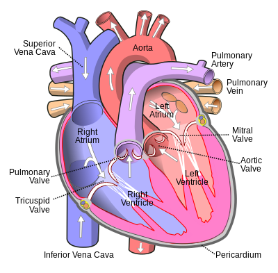
The Heart: A Double Pump
The heart is a muscular organ about the size of a fist, located in the mediastinum between the lungs. It has four chambers: two atria (upper receiving chambers) and two ventricles (lower pumping chambers). The right side pumps deoxygenated blood to the lungs (pulmonary circulation); the left side pumps oxygenated blood to the body (systemic circulation). The heart beats about 100,000 times per day, pumping approximately 7,000 liters of blood.
Heart Valves & Blood Flow
Four valves ensure one-directional blood flow: the tricuspid valve (right atrium to right ventricle), pulmonary valve (right ventricle to pulmonary arteries), mitral/bicuspid valve (left atrium to left ventricle), and aortic valve (left ventricle to aorta). Heart sounds ('lub-dub') result from valve closure. The cardiac cycle includes diastole (relaxation/filling) and systole (contraction/ejection). Normal blood pressure is approximately 120/80 mmHg.
The Electrical Conduction System
The heart has its own electrical system generating and conducting impulses. The SA node (sinoatrial node) in the right atrium is the natural pacemaker, firing 60-100 times per minute. The impulse travels to the AV node (atrioventricular), then down the Bundle of His, left and right bundle branches, to the Purkinje fibers — coordinating atrial and ventricular contractions. An electrocardiogram (ECG/EKG) records this electrical activity.
Blood Vessels
Arteries carry blood away from the heart under high pressure — they have thick, elastic walls with smooth muscle. Arterioles regulate blood flow to capillaries. Capillaries are microscopic one-cell-thick vessels where oxygen, nutrients, and wastes are exchanged between blood and tissues. Venules collect capillary blood into veins, which carry blood back to the heart under low pressure and have valves to prevent backflow.
Blood Composition
Blood is a liquid connective tissue composing about 8% of body weight (~5-6 liters). It consists of plasma (55% — water, proteins, hormones, nutrients, wastes) and formed elements (45%): erythrocytes/red blood cells (carry oxygen via hemoglobin, live 120 days), leukocytes/white blood cells (immune defense, 5 types), and thrombocytes/platelets (blood clotting). The ABO blood typing system identifies four major blood groups: A, B, AB, and O.
The Respiratory System
Breathing & Gas Exchange

Upper Respiratory Tract
Air enters through the nose and mouth. The nasal cavity is lined with mucosa and cilia that filter, warm, and humidify air. The paranasal sinuses (frontal, maxillary, ethmoid, sphenoid) lighten the skull and resonate the voice. The pharynx (throat) is shared by respiratory and digestive systems. The larynx (voice box) contains the vocal cords and the epiglottis — a cartilage flap that prevents food from entering the airway during swallowing.
Lower Respiratory Tract
The trachea (windpipe) is reinforced by C-shaped cartilage rings, extending from the larynx to the primary bronchi. It bifurcates at the carina into the right and left primary bronchi, which enter the lungs and branch progressively: lobar bronchi → segmental bronchi → bronchioles → terminal bronchioles → respiratory bronchioles → alveolar ducts → alveoli. This branching creates the 'bronchial tree.' The right lung has 3 lobes; the left has 2 (to accommodate the heart).
Alveoli: Site of Gas Exchange
The lungs contain approximately 300 million alveoli, providing a total surface area of 70 m² — the size of a tennis court! Each alveolus is surrounded by pulmonary capillaries. Oxygen diffuses from alveoli into blood; carbon dioxide diffuses from blood into alveoli — driven by concentration gradients. Alveolar walls are only one cell thick. Type II pneumocytes produce surfactant, a lipoprotein that reduces surface tension and prevents alveolar collapse.
Mechanics of Breathing
Breathing is a mechanical process driven by pressure changes. During inspiration, the diaphragm contracts and flattens, external intercostals lift the ribs, chest volume increases, pressure drops below atmospheric, and air flows in. During quiet expiration, these muscles relax, chest recoils, pressure rises, and air flows out. At rest, the tidal volume is ~500 mL per breath; total lung capacity is about 6 liters. The respiratory rate is 12–20 breaths per minute.
Control of Respiration
Breathing is controlled by the respiratory center in the medulla oblongata and pons. Chemoreceptors monitor blood CO₂, O₂, and pH levels. Rising CO₂ (acidosis) is the primary stimulus to breathe — it triggers faster, deeper breaths. The Hering-Breuer reflex prevents overinflation of the lungs. We can voluntarily control breathing (holding breath, speaking), but if CO₂ rises too high, involuntary control overrides the voluntary effort.
The Digestive System
Nutrition, Digestion & Absorption

Overview: The Alimentary Canal
The digestive system processes food through a tube approximately 9 meters long — the alimentary canal (gastrointestinal tract) — plus accessory organs. Six processes occur: ingestion (eating), propulsion (swallowing and peristalsis), mechanical digestion (chewing, churning), chemical digestion (enzymes breaking down food), absorption (nutrients into blood/lymph), and defecation (eliminating waste). The entire journey from mouth to anus takes 24–72 hours.
Mouth, Pharynx & Esophagus
Digestion begins in the mouth with mastication (chewing) and salivary amylase breaking down starch. The tongue shapes food into a bolus and initiates swallowing (deglutition). The bolus passes through the pharynx into the esophagus — a muscular tube ~25 cm long. Peristalsis — rhythmic waves of muscular contraction — propels food to the stomach in about 8 seconds. The lower esophageal sphincter prevents stomach acid reflux.
The Stomach
The J-shaped stomach holds up to 4 liters and performs both mechanical (churning) and chemical digestion. Gastric glands secrete: hydrochloric acid (HCl, pH 1.5–3.5 — kills bacteria and activates enzymes), pepsinogen (converted to pepsin, which digests proteins), intrinsic factor (essential for vitamin B12 absorption), and mucus (protects stomach wall). After 2–6 hours, chyme (semifluid mixture) passes into the small intestine through the pyloric sphincter.
Small Intestine: The Absorption Powerhouse
The small intestine (~6 meters long, 2.5 cm diameter) is divided into the duodenum (receives bile and pancreatic enzymes), jejunum, and ileum. The intestinal wall has structural adaptations maximizing surface area to 200 m²: circular folds (plicae circulares), finger-like projections (villi), and tiny projections on each cell (microvilli/brush border). 90% of nutrient absorption occurs here — sugars and amino acids into capillaries; fats into lymph vessels (lacteals).
Large Intestine, Liver & Pancreas
The large intestine (~1.5 m long, 6.5 cm wide) includes cecum, colon (ascending, transverse, descending, sigmoid), rectum, and anal canal. Its main function is water absorption — converting liquid chyme to solid feces. The liver (largest internal organ, ~1.5 kg) produces bile for fat emulsification, detoxifies blood, and performs 500+ functions. The pancreas produces digestive enzymes (lipase, amylase, proteases) and the hormones insulin and glucagon.
The Endocrine System
Hormones & Chemical Signaling
Hormones & Glands
The endocrine system communicates through hormones — chemical messengers secreted into the bloodstream that act on distant target organs. Unlike the nervous system (milliseconds), hormonal responses take seconds to hours but last longer. Endocrine glands include the pituitary, thyroid, parathyroid, adrenal, pancreas, gonads (testes/ovaries), pineal, and thymus. Hormones are classified as amino acid-based (water-soluble, bind surface receptors) or steroid (lipid-soluble, enter cells).
The Pituitary: The Master Gland
The pituitary gland (size of a pea) sits in the sella turcica of the sphenoid bone and is controlled by the hypothalamus. The anterior pituitary produces: GH (growth hormone), TSH (thyroid-stimulating), ACTH (adrenocorticotropic), FSH and LH (gonadotropins), and prolactin. The posterior pituitary releases ADH (antidiuretic hormone, regulates water balance) and oxytocin (uterine contractions, milk ejection, social bonding). This one gland influences virtually every other hormone in the body.
Thyroid & Adrenal Glands
The thyroid gland (butterfly-shaped, in neck) produces T3 and T4 (thyroid hormones that regulate metabolic rate) and calcitonin (lowers blood calcium). Hypothyroidism causes fatigue, weight gain, and cold intolerance; hyperthyroidism causes weight loss and rapid heartbeat. The adrenal glands sit atop each kidney: the cortex produces cortisol (stress response, anti-inflammatory), aldosterone (sodium/potassium balance), and androgens; the medulla releases epinephrine and norepinephrine (fight-or-flight).
Pancreatic Hormones & Blood Glucose
The pancreatic islets of Langerhans produce insulin (beta cells — released when blood glucose is high, promotes glucose uptake into cells and glycogen storage) and glucagon (alpha cells — released when blood glucose is low, stimulates glycogenolysis and gluconeogenesis in the liver). Type 1 diabetes results from autoimmune destruction of beta cells; Type 2 from insulin resistance. Approximately 537 million adults worldwide have diabetes.
The Reproductive System
Continuation of Life

The Heart: A Double Pump
The heart is a muscular organ about the size of a fist, located in the mediastinum between the lungs. It has four chambers: two atria (upper receiving chambers) and two ventricles (lower pumping chambers). The right side pumps deoxygenated blood to the lungs (pulmonary circulation); the left side pumps oxygenated blood to the body (systemic circulation). The heart beats about 100,000 times per day, pumping approximately 7,000 liters of blood.
Heart Valves & Blood Flow
Four valves ensure one-directional blood flow: the tricuspid valve (right atrium to right ventricle), pulmonary valve (right ventricle to pulmonary arteries), mitral/bicuspid valve (left atrium to left ventricle), and aortic valve (left ventricle to aorta). Heart sounds ('lub-dub') result from valve closure. The cardiac cycle includes diastole (relaxation/filling) and systole (contraction/ejection). Normal blood pressure is approximately 120/80 mmHg.
The Electrical Conduction System
The heart has its own electrical system generating and conducting impulses. The SA node (sinoatrial node) in the right atrium is the natural pacemaker, firing 60-100 times per minute. The impulse travels to the AV node (atrioventricular), then down the Bundle of His, left and right bundle branches, to the Purkinje fibers — coordinating atrial and ventricular contractions. An electrocardiogram (ECG/EKG) records this electrical activity.
Blood Vessels
Arteries carry blood away from the heart under high pressure — they have thick, elastic walls with smooth muscle. Arterioles regulate blood flow to capillaries. Capillaries are microscopic one-cell-thick vessels where oxygen, nutrients, and wastes are exchanged between blood and tissues. Venules collect capillary blood into veins, which carry blood back to the heart under low pressure and have valves to prevent backflow.
Blood Composition
Blood is a liquid connective tissue composing about 8% of body weight (~5-6 liters). It consists of plasma (55% — water, proteins, hormones, nutrients, wastes) and formed elements (45%): erythrocytes/red blood cells (carry oxygen via hemoglobin, live 120 days), leukocytes/white blood cells (immune defense, 5 types), and thrombocytes/platelets (blood clotting). The ABO blood typing system identifies four major blood groups: A, B, AB, and O.
The Respiratory System
Breathing & Gas Exchange
Upper Respiratory Tract
Air enters through the nose and mouth. The nasal cavity is lined with mucosa and cilia that filter, warm, and humidify air. The paranasal sinuses (frontal, maxillary, ethmoid, sphenoid) lighten the skull and resonate the voice. The pharynx (throat) is shared by respiratory and digestive systems. The larynx (voice box) contains the vocal cords and the epiglottis — a cartilage flap that prevents food from entering the airway during swallowing.
Lower Respiratory Tract
The trachea (windpipe) is reinforced by C-shaped cartilage rings, extending from the larynx to the primary bronchi. It bifurcates at the carina into the right and left primary bronchi, which enter the lungs and branch progressively: lobar bronchi → segmental bronchi → bronchioles → terminal bronchioles → respiratory bronchioles → alveolar ducts → alveoli. This branching creates the 'bronchial tree.' The right lung has 3 lobes; the left has 2 (to accommodate the heart).
Alveoli: Site of Gas Exchange
The lungs contain approximately 300 million alveoli, providing a total surface area of 70 m² — the size of a tennis court! Each alveolus is surrounded by pulmonary capillaries. Oxygen diffuses from alveoli into blood; carbon dioxide diffuses from blood into alveoli — driven by concentration gradients. Alveolar walls are only one cell thick. Type II pneumocytes produce surfactant, a lipoprotein that reduces surface tension and prevents alveolar collapse.
Mechanics of Breathing
Breathing is a mechanical process driven by pressure changes. During inspiration, the diaphragm contracts and flattens, external intercostals lift the ribs, chest volume increases, pressure drops below atmospheric, and air flows in. During quiet expiration, these muscles relax, chest recoils, pressure rises, and air flows out. At rest, the tidal volume is ~500 mL per breath; total lung capacity is about 6 liters. The respiratory rate is 12–20 breaths per minute.
Control of Respiration
Breathing is controlled by the respiratory center in the medulla oblongata and pons. Chemoreceptors monitor blood CO₂, O₂, and pH levels. Rising CO₂ (acidosis) is the primary stimulus to breathe — it triggers faster, deeper breaths. The Hering-Breuer reflex prevents overinflation of the lungs. We can voluntarily control breathing (holding breath, speaking), but if CO₂ rises too high, involuntary control overrides the voluntary effort.
The Digestive System
Nutrition, Digestion & Absorption
Overview: The Alimentary Canal
The digestive system processes food through a tube approximately 9 meters long — the alimentary canal (gastrointestinal tract) — plus accessory organs. Six processes occur: ingestion (eating), propulsion (swallowing and peristalsis), mechanical digestion (chewing, churning), chemical digestion (enzymes breaking down food), absorption (nutrients into blood/lymph), and defecation (eliminating waste). The entire journey from mouth to anus takes 24–72 hours.
Mouth, Pharynx & Esophagus
Digestion begins in the mouth with mastication (chewing) and salivary amylase breaking down starch. The tongue shapes food into a bolus and initiates swallowing (deglutition). The bolus passes through the pharynx into the esophagus — a muscular tube ~25 cm long. Peristalsis — rhythmic waves of muscular contraction — propels food to the stomach in about 8 seconds. The lower esophageal sphincter prevents stomach acid reflux.
The Stomach
The J-shaped stomach holds up to 4 liters and performs both mechanical (churning) and chemical digestion. Gastric glands secrete: hydrochloric acid (HCl, pH 1.5–3.5 — kills bacteria and activates enzymes), pepsinogen (converted to pepsin, which digests proteins), intrinsic factor (essential for vitamin B12 absorption), and mucus (protects stomach wall). After 2–6 hours, chyme (semifluid mixture) passes into the small intestine through the pyloric sphincter.
Small Intestine: The Absorption Powerhouse
The small intestine (~6 meters long, 2.5 cm diameter) is divided into the duodenum (receives bile and pancreatic enzymes), jejunum, and ileum. The intestinal wall has structural adaptations maximizing surface area to 200 m²: circular folds (plicae circulares), finger-like projections (villi), and tiny projections on each cell (microvilli/brush border). 90% of nutrient absorption occurs here — sugars and amino acids into capillaries; fats into lymph vessels (lacteals).
Large Intestine, Liver & Pancreas
The large intestine (~1.5 m long, 6.5 cm wide) includes cecum, colon (ascending, transverse, descending, sigmoid), rectum, and anal canal. Its main function is water absorption — converting liquid chyme to solid feces. The liver (largest internal organ, ~1.5 kg) produces bile for fat emulsification, detoxifies blood, and performs 500+ functions. The pancreas produces digestive enzymes (lipase, amylase, proteases) and the hormones insulin and glucagon.
The Endocrine System
Hormones & Chemical Signaling
Hormones & Glands
The endocrine system communicates through hormones — chemical messengers secreted into the bloodstream that act on distant target organs. Unlike the nervous system (milliseconds), hormonal responses take seconds to hours but last longer. Endocrine glands include the pituitary, thyroid, parathyroid, adrenal, pancreas, gonads (testes/ovaries), pineal, and thymus. Hormones are classified as amino acid-based (water-soluble, bind surface receptors) or steroid (lipid-soluble, enter cells).
The Pituitary: The Master Gland
The pituitary gland (size of a pea) sits in the sella turcica of the sphenoid bone and is controlled by the hypothalamus. The anterior pituitary produces: GH (growth hormone), TSH (thyroid-stimulating), ACTH (adrenocorticotropic), FSH and LH (gonadotropins), and prolactin. The posterior pituitary releases ADH (antidiuretic hormone, regulates water balance) and oxytocin (uterine contractions, milk ejection, social bonding). This one gland influences virtually every other hormone in the body.
Thyroid & Adrenal Glands
The thyroid gland (butterfly-shaped, in neck) produces T3 and T4 (thyroid hormones that regulate metabolic rate) and calcitonin (lowers blood calcium). Hypothyroidism causes fatigue, weight gain, and cold intolerance; hyperthyroidism causes weight loss and rapid heartbeat. The adrenal glands sit atop each kidney: the cortex produces cortisol (stress response, anti-inflammatory), aldosterone (sodium/potassium balance), and androgens; the medulla releases epinephrine and norepinephrine (fight-or-flight).
Pancreatic Hormones & Blood Glucose
The pancreatic islets of Langerhans produce insulin (beta cells — released when blood glucose is high, promotes glucose uptake into cells and glycogen storage) and glucagon (alpha cells — released when blood glucose is low, stimulates glycogenolysis and gluconeogenesis in the liver). Type 1 diabetes results from autoimmune destruction of beta cells; Type 2 from insulin resistance. Approximately 537 million adults worldwide have diabetes.
The Reproductive System
Continuation of Life
Male Reproductive System
The male reproductive system produces, stores, and delivers sperm. The testes (in the scrotum, 2–3°C below body temperature — optimal for spermatogenesis) produce sperm and testosterone. Sperm mature in the epididymis, travel through the vas deferens, mix with secretions from the seminal vesicles (fructose-rich fluid), prostate gland (alkaline fluid), and bulbourethral glands to form semen. Each ejaculate contains 200–500 million sperm. Sperm take ~74 days to develop.
Female Reproductive System
The female reproductive system produces eggs (ova) and provides the environment for fertilization and fetal development. The ovaries produce oocytes and hormones (estrogen, progesterone). Each month, follicle-stimulating hormone (FSH) triggers follicle maturation; a surge of LH causes ovulation (release of the oocyte). The fallopian tubes transport the egg toward the uterus — fertilization typically occurs in the ampulla. The uterus (pear-shaped muscular organ) nurtures the developing embryo; the cervix is its narrow inferior opening.
The Menstrual Cycle
The menstrual cycle averages 28 days: Days 1–5 (menstruation — shedding of endometrium), Days 6–13 (follicular phase — rising estrogen thickens endometrium), Day 14 (ovulation — LH surge), Days 15–28 (luteal phase — corpus luteum produces progesterone maintaining endometrium). If fertilization doesn't occur, progesterone drops, triggering menstruation. The cycle continues from menarche (~age 12) to menopause (~age 51).
Fertilization & Early Development
Fertilization occurs when one sperm penetrates the secondary oocyte, forming a zygote (diploid, 46 chromosomes). The zygote undergoes cleavage as it travels to the uterus (~3–4 days), forming the morula, then blastocyst. Implantation occurs 6–10 days after fertilization. The inner cell mass develops into the embryo; the outer trophoblast forms the placenta — which provides oxygen and nutrients, removes wastes, and produces hCG (detected by pregnancy tests), estrogen, and progesterone.
The Integumentary System
Skin, Hair & Nails

Skin: The Largest Organ
The skin is the body's largest organ, covering about 1.8 m² and weighing 4–5 kg. It has three main layers: the epidermis (outer, avascular), dermis (inner, thick, vascular), and hypodermis/subcutaneous tissue (beneath the dermis, mainly fat). Skin color is determined by melanin (produced by melanocytes), carotene, and hemoglobin. Skin functions as a physical barrier, thermoregulator, sensory organ, vitamin D synthesizer, and immune defense.
The Epidermis
The epidermis is stratified squamous epithelium with five layers (strata): stratum basale (deepest — mitotically active stem cells and melanocytes), stratum spinosum, stratum granulosum, stratum lucidum (only in thick skin of palms and soles), and stratum corneum (outermost — 20–30 layers of dead, keratin-filled cells). Keratinocytes journey from basale to corneum in 25–45 days. Langerhans cells in the epidermis are immune sentinels against pathogens.
The Dermis & Its Structures
The dermis contains collagen and elastic fibers (providing strength and elasticity), blood vessels, nerve endings (touch, pain, temperature receptors), hair follicles, sebaceous (oil) glands, sudoriferous (sweat) glands — eccrine (temperature regulation) and apocrine (stress/emotional sweating) — and arrector pili muscles (cause goosebumps). Fingerprints arise from dermal ridges — unique to each individual, used in forensics for over a century.
Hair, Nails & Wound Healing
Hair is composed of dead keratinized cells; the follicle is the living root where growth occurs. Hair protects, senses, and insulates. The hair cycle includes anagen (growth, 2–7 years), catagen (transition), and telogen (resting/shedding, ~100 hairs/day). Nails are hardened keratin plates protecting fingertips and aiding grip. Wound healing occurs in four overlapping phases: hemostasis (clot formation), inflammation, proliferation (new tissue), and remodeling (scar maturation).
The Urinary System
Filtration, Excretion & Homeostasis

The Kidneys: Master Filters
The two kidneys (bean-shaped, ~11 cm) sit retroperitoneally at T12–L3. Each kidney contains ~1 million nephrons — the functional filtration units. Every day, the kidneys filter ~180 liters of blood plasma, reabsorb 99% of it, and produce ~1–2 liters of urine. Kidneys regulate blood pressure (via renin-angiotensin-aldosterone system), red blood cell production (erythropoietin), blood pH, blood volume, and electrolyte balance (sodium, potassium, calcium, phosphate).
The Nephron: Structure & Function
Each nephron consists of: glomerulus (capillary knot where filtration occurs under pressure), Bowman's capsule (collects filtrate), proximal convoluted tubule (reabsorbs 65% of water, glucose, amino acids, ions), loop of Henle (creates osmotic gradient for water reabsorption), distal convoluted tubule (fine-tunes ion balance under hormonal control), and collecting duct (final water reabsorption regulated by ADH). The processes are filtration, reabsorption, secretion, and excretion.
Urine Formation & Composition
Urine is typically light yellow (due to urochrome pigment from hemoglobin breakdown), slightly acidic (pH 4.5–8.0), with a specific gravity of 1.001–1.035. Normal urine contains water (95%), urea (main nitrogenous waste from protein metabolism), creatinine, uric acid, ions, and trace hormones. Abnormal findings include glucose (diabetes mellitus), proteins (kidney disease), blood (hematuria), ketones (diabetic ketoacidosis), and bilirubin (liver disease).
Bladder, Ureters & Urethra
Urine drains from each renal pelvis through ureters (25–30 cm muscular tubes) to the urinary bladder — a hollow, distensible muscular sac capable of holding 500–600 mL. The bladder wall has the detrusor muscle and a trigone (triangular area between ureteral openings and urethral outlet). The internal urethral sphincter (involuntary) and external urethral sphincter (voluntary) control micturition. The male urethra is ~20 cm; female urethra is ~4 cm (explaining higher UTI frequency in females).
The Lymphatic & Immune System
Defense Against Disease

The Lymphatic System
The lymphatic system consists of lymphatic vessels, lymph nodes, and lymphoid organs (spleen, thymus, tonsils, MALT). Lymphatic capillaries collect excess interstitial fluid (plasma that leaked from capillaries) and return it to the bloodstream — preventing edema. This fluid, once in lymphatics, is called lymph. Lymph travels through progressively larger vessels, passes through lymph nodes (where it is filtered), and empties into the subclavian veins. The thoracic duct is the largest lymphatic vessel.
Innate Immunity: The First Line of Defense
Innate immunity provides rapid, non-specific protection. Physical barriers include skin and mucous membranes. Chemical barriers include stomach acid, lysozyme in tears, and defensins in mucus. Cellular defenses include phagocytes (neutrophils and macrophages that engulf pathogens), natural killer (NK) cells (destroy abnormal cells), and dendritic cells. The inflammatory response — redness, heat, swelling, pain — is triggered by histamine and cytokines, recruiting immune cells to infected areas.
Adaptive Immunity: Targeted Defense
Adaptive immunity is specific, has immunological memory, and takes 1–2 weeks to fully activate. B lymphocytes produce antibodies (immunoglobulins) that bind specific antigens — this is humoral immunity. T lymphocytes include helper T cells (coordinate the immune response), cytotoxic T cells (kill infected or cancerous cells), and regulatory T cells (prevent autoimmunity) — this is cell-mediated immunity. Memory B and T cells persist after infection, enabling rapid responses upon re-exposure.
Vaccines & Immunological Memory
Vaccines exploit immunological memory by exposing the immune system to a harmless form of an antigen (weakened pathogen, inactivated toxin, or mRNA instructions). The primary immune response is slow; memory cells are formed. Upon re-exposure, the secondary immune response is faster, stronger, and longer-lasting — often preventing disease before symptoms appear. Edward Jenner's 1796 smallpox vaccine was the first; vaccination has since eradicated smallpox and nearly eliminated polio globally.